It’s nine years since an enigmatic disease emerged and began destroying the region’s coral reef system, and scientists still cannot identify the pathogen driving the contagion, but a recent study may move them a little closer to fingering the mysterious killer.
The authors of the study describe Stony Coral Tissue Loss Disease as “the most pervasive and contagious coral disease on record.”
First spotted off the coast of Florida in 2014, the fast-moving SCTLD has killed and damaged much of the coral along the 350 miles of the state’s famous Coral Reef. By 2018 it had made its way to the Caribbean and it was first seen in V.I. waters in January 2019.
It is now documented in waters off 22 Caribbean islands, including all of the Virgin Islands and Puerto Rico. It has proven deadly to more than 20 different species of coral.
Efforts to counter the disease have included limited success with antibiotics. But the real key to defeating SCTLD lies in understanding what causes it and how it progresses.
That was the motivation for a major research project led by Marilyn Brandt, research professor of marine science at the University of the Virgin Islands, and Laura Mydlarz, professor of biology at the University of Texas at Arlington. Scientists from Mote Marine Laboratory, Woods Hole Oceanographic Institution, Louisiana State University and Rice University also collaborated on the study.

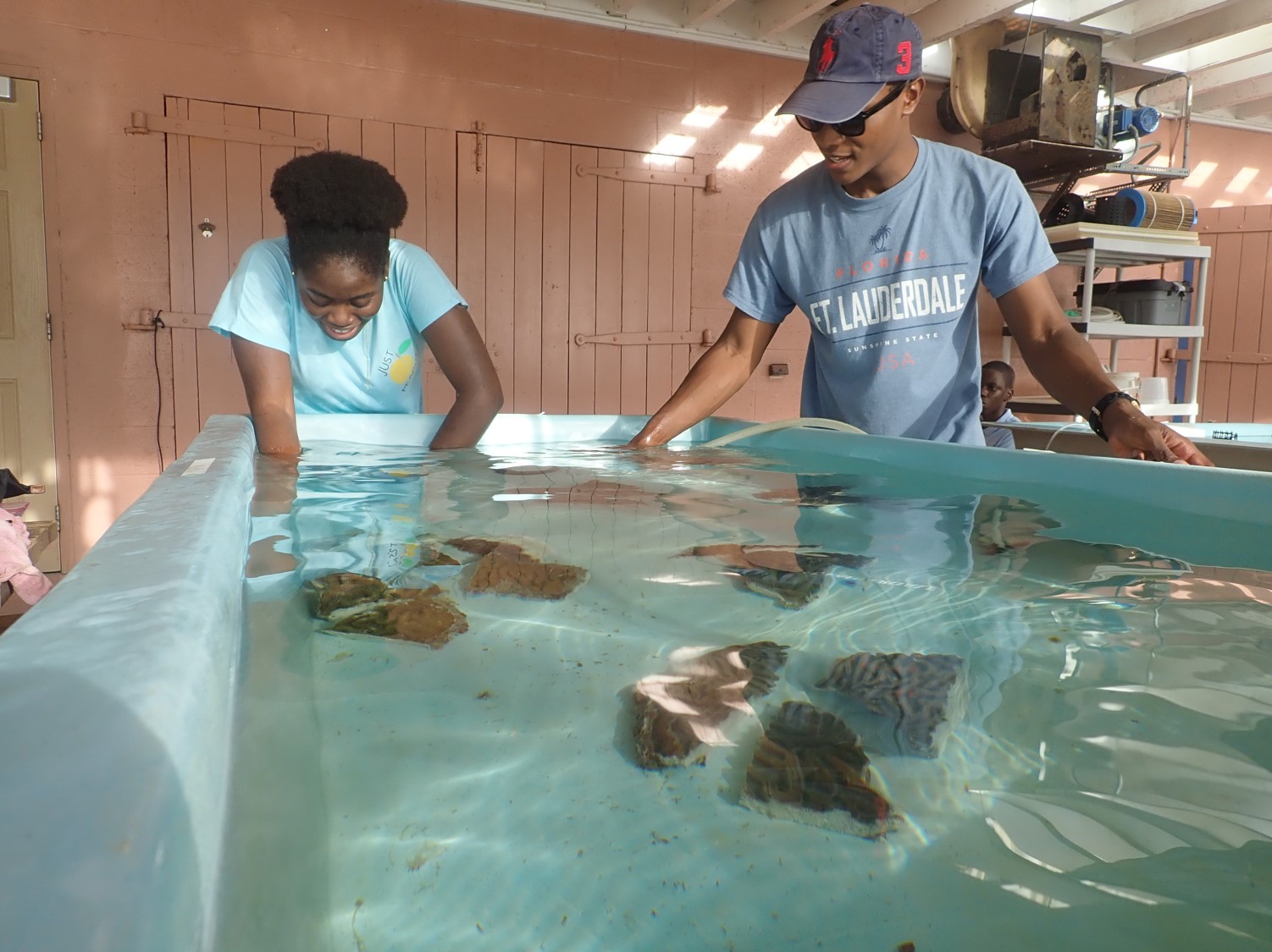
The Marine Science Center at the University of the Virgin Islands was the site of experimental research on the transmission of Stony Coral Tissue Loss Disease. (Photo by Sonora Meiling)
Brandt’s team at UVI conducted a transmission experiment, tracing how the disease spreads from infected to healthy coral. Researchers at UTA examined the gene structure and variants of the affected coral.
“We wanted to compare how the different species reacted to the disease, thinking that if we could find commonalities among the responses, it would give us clues to how the disease works and maybe even help to identify a pathogenic agent,” Brandt said.
They exposed samples of five distinct species of coral to SCTLD. What they observed supports the theory that the pathogen is a virus. Corals developing visible lesions from SCTLD activated an antiviral immune response.
Although the study was far from conclusive, the suggestion that the pathogen is some sort of virus is consistent with “multiple lines of evidence” from other studies, the report states.
The UVI and UTA researchers also found that the pathogen interrupts the natural relationship between coral and Symbiodinaceas, an algae that lives in coral in a symbiotic relationship.
Under normal conditions, the coral provides a safe environment for the algae, and the algae, through photosynthesis, provide food for the coral. The algae are “fundamental” to the coral’s survival, Brandt said. SCTLD can infect both the coral and the algae.
The study showed that the coral reacted to infected Symbiodinaceas by increasing a particular gene, rab7, which is found in coral that digests dead or dysfunctional symbiotic partners.
The research also provided a plausible explanation for why antibiotic substances applied to infected coral are able to arrest existing SCTLD lesions (they are 84 percent effective) yet cannot prevent new lesions from appearing on other parts of the same coral colony.
The lesions may be an indirect result of the disease. Corals have a protective mucus layer that defends them against foreign particles and microbes. SCTLD appears to render corals incapable of maintaining the mucus layer. Without that layer, they become susceptible to “secondary infection by opportunistic bacteria,” the report says.
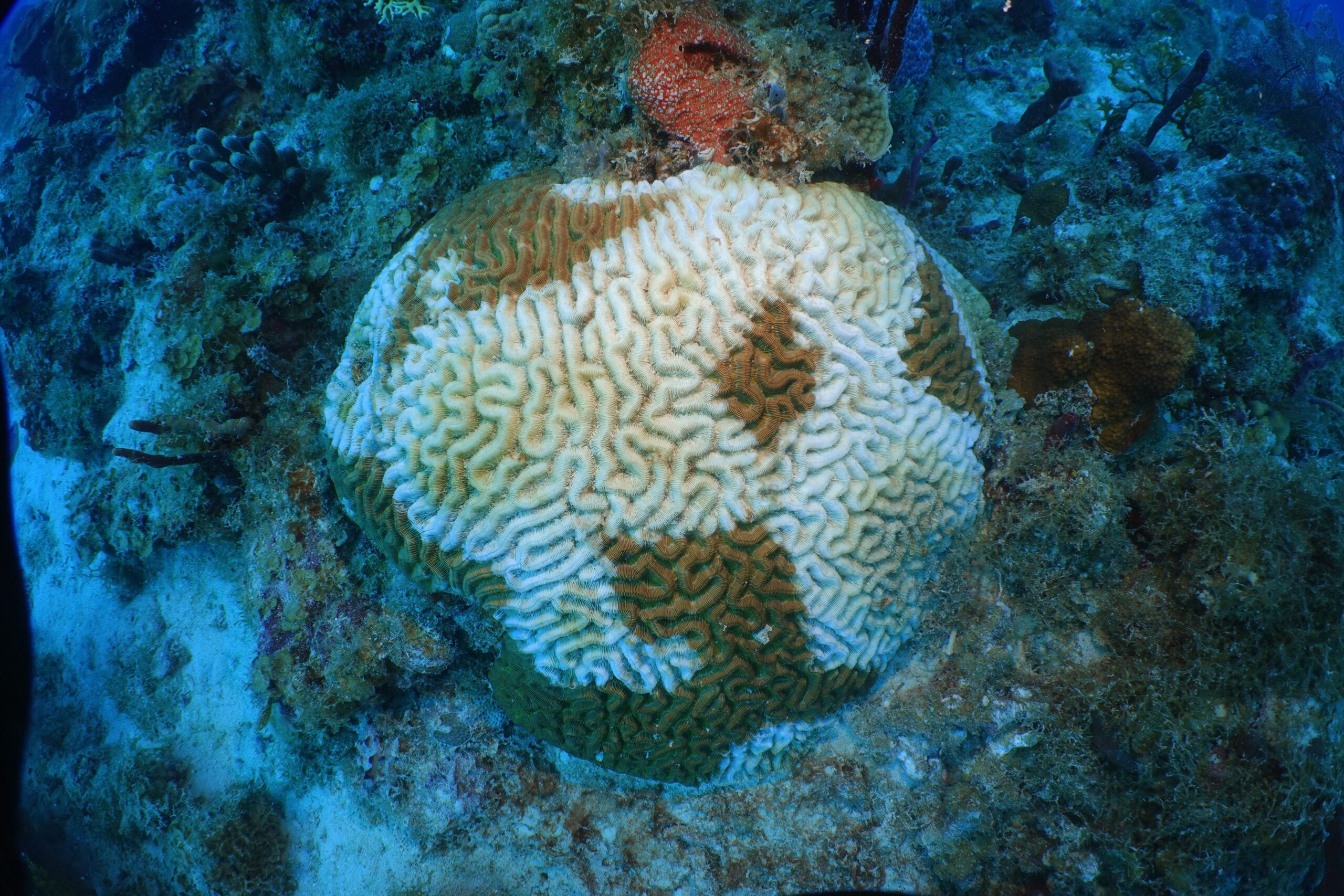
The study also documented the already generally held belief that some species of coral are more susceptible to SCTLD than others. Of the five species in the experiment, the two most dramatically affected were boulder brain coral (also called large-grooved brain coral) and boulder star coral.
Numerous projects are already underway to try to grow these and other corals and to “plant” them in efforts to restore reefs.
SCTLD may be the worst yet, but it is just the most recent in a series of phenomena affecting the ocean’s coral reefs, including numerous massive bleaching events blamed at least in part on unusually warm waters resulting from global warming.
Since the 1970s, the report states, there has been a “decline in living coral tissue” of between 50 percent and 80 percent.
“As climate change pressures continue to escalate, emerging and endemic outbreaks in the Caribbean are likely to decimate reef-building coral populations, resulting in reef ecosystem collapse,” the report states.
A sense of urgency is not lost on the scientists studying the problems. Coral research, including on SCTLD, is ongoing on several fronts, including at Brandt’s UVI lab, where researchers are working now with samples they took last year from wild corals.
Stony Coral Tissue Loss Disease could be the result of a cumulation of pressures. There were unprecedented back-to-back coral bleaching events in Florida waters in 2014 and 2015, Brandt said. At the same time, there was a major, deep-water dredging project that, some theorize, could have disturbed and distributed a previously unknown pathogen.
“New pathogens emerge all the time,” she said.
So far, the identity of the one causing SCTLD still eludes scientists.
“We’ve looked and looked,” Brandt said, but “there’s no smoking gun. . . We’re working as hard as we can and as fast as we can to get this information.”
The study, “Stony coral tissue loss disease induces transcriptional signatures of in situ degradation of dysfunctional Symbiodinaceae,” was published in the May 22 edition of Nature Communications.

